Home
/ Neck And Upper Back Anatomy : Anatomy Of The Back Spine And Back Muscles Kenhub, Tackle it to learn more about the bones, vessels, muscles and organs of the head and neck!
Neck And Upper Back Anatomy : Anatomy Of The Back Spine And Back Muscles Kenhub, Tackle it to learn more about the bones, vessels, muscles and organs of the head and neck!
Neck And Upper Back Anatomy : Anatomy Of The Back Spine And Back Muscles Kenhub, Tackle it to learn more about the bones, vessels, muscles and organs of the head and neck!. Anatomy of the chest and stomach 12 photos of the anatomy of the chest and stomach anatomy of the chest and stomach, human anatomy, anatomy of the chest and stomach. The rib cage also anchors the bones of the head, neck, shoulders, and arms to the trunk of the body. Neck anatomy nerves picture there are 8 spinal nerves that originate from the cervical spine. The top of the cervical spine connects to the skull, and the bottom connects to the upper back at about shoulder level. These important muscles control many motions that involve moving the arms and head — such as throwing a ball, looking up at the sky, and raising your hand.
The cervical area consists of 7 vertebrae in the neck. The phrenic nerve originates in the cervical spinal nerves of the neck, but descends through the thorax to innervate the thoracic diaphragm. The muscles of the chest and upper back occupy the thoracic region of the body inferior to the neck and superior to the abdominal region and include the muscles of the shoulders. The sacrum has 5 small, fused vertebrae. The atlas (c1) and axis (c2) are part of the spine's craniovertebral junction (cvj)—this is where the base of your brain becomes part of your spinal column.
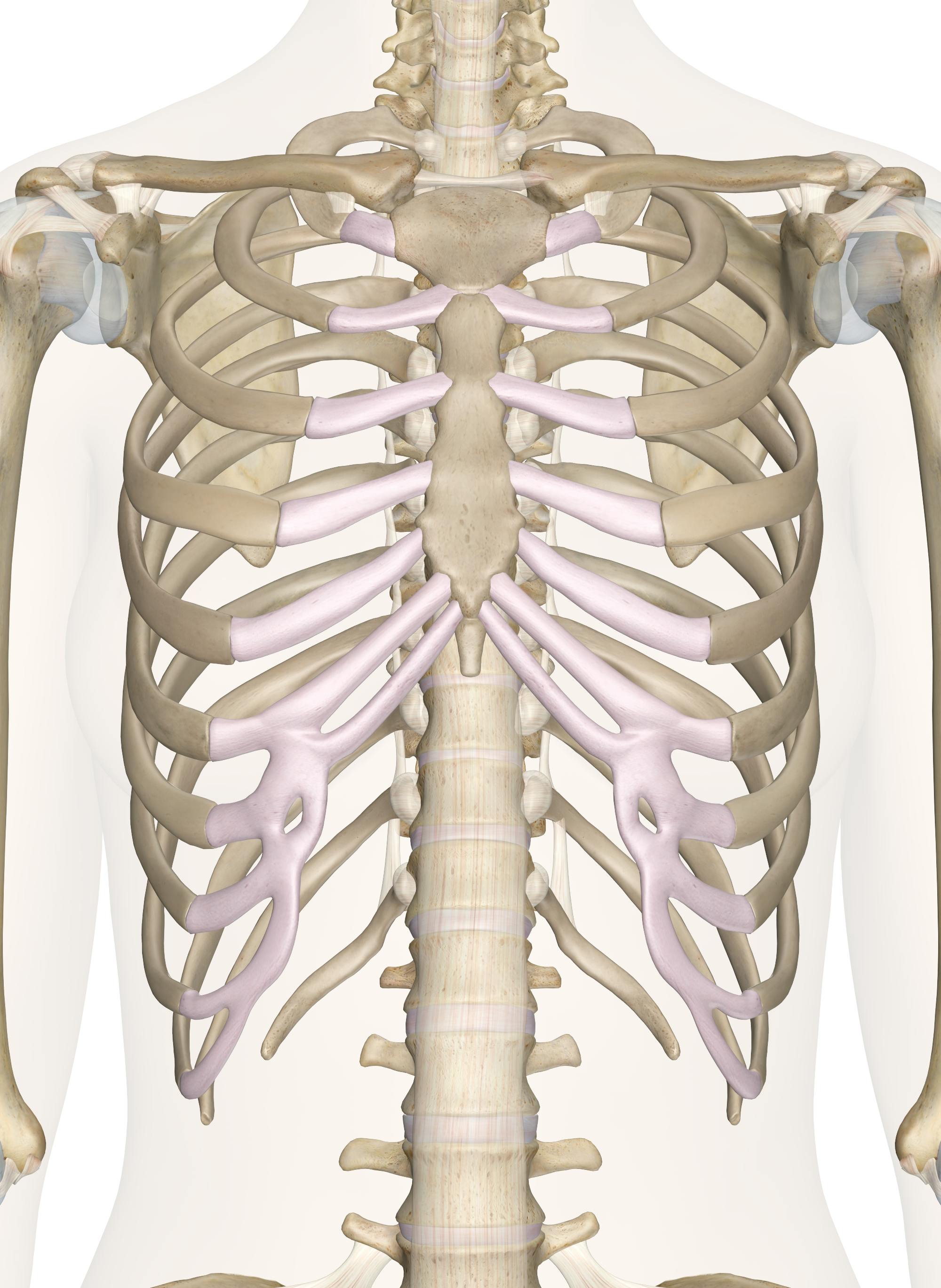
Bones Of The Chest And Upper Back from innerbody.imgix.net The content of the neck is grouped into 4 neck spaces, called the compartments. The thoracic area consists of 12 vertebrae in the chest. The upper trapezius arises from your occipital bone in the back of your skull and the nuchal line in the back of your neck. Discs lose some of their. The muscles of the back muscles make up a large part of the anatomy (structure) of the back. The efferent signals of the phrenic nerve cause the contractions of the diaphragm that permit breathing and keep the body alive. The cervical spine supports the weight and movement of your head and protects the nerves exiting your brain. The back is the body region between the neck and the gluteal regions.
This is my video about the muscles of the back.
The phrenic nerve originates in the cervical spinal nerves of the neck, but descends through the thorax to innervate the thoracic diaphragm. Contains glands ( thyroid, parathyroid, and thymus ), the larynx, pharynx and trachea. Causes of neck pain and how to manage the pain in basic terms, the neck (cervical spine) joins the shoulders and chest to the head. The top of the cervical spine connects to the skull, and the bottom connects to the upper back at about shoulder level. It is made up of bones, discs, muscles, ligaments, nerves and tendons. This is my video about the muscles of the back. They start at the top of the neck and go down to the tailbone. The seventh cervical vertebra is located at the top of your shoulder and upper back. The thoracic area consists of 12 vertebrae in the chest. Contains cervical vertebrae and postural muscles. Both the deltoid and the trapezius are firmly attached to the spine of the scapula. The back is the body region between the neck and the gluteal regions. These important muscles control many motions that involve moving the arms and head — such as throwing a ball, looking up at the sky, and raising your hand.
The atlas (c1) and axis (c2) are part of the spine's craniovertebral junction (cvj)—this is where the base of your brain becomes part of your spinal column. The cervical spine is responsible for several crucial roles, including. This is my video about the muscles of the back. The majority of these nerves control the functions of the upper extremities and allow you to feel your arms, shoulder, and back of your head. It comprises the vertebral column (spine) and two compartments of back muscles;
The Extreme Dangers Of Ignoring Upper Back And Neck Pain Longmont Spine Center from www.longmontspinecenter.com The thoracic area consists of 12 vertebrae in the chest. The sacrum has 5 small, fused vertebrae. Pain and dysfunction from injuries or conditions that impact the joints, muscles, and other structures can easily spread from the neck to the shoulder (s) and from the shoulder (s) to the neck. The neck begins at the base of the skull and connects to the thoracic spine (the upper back). The lumbar area consists of 5 vertebrae in the lower back. The content of the neck is grouped into 4 neck spaces, called the compartments. It is made up of bones, discs, muscles, ligaments, nerves and tendons. Contains glands ( thyroid, parathyroid, and thymus ), the larynx, pharynx and trachea.
The lateral neck muscles, also called the lateral vertebral muscles, are a group of muscles that pass obliquely along the lateral sides of the neck.
It comprises the vertebral column (spine) and two compartments of back muscles; The sacrum has 5 small, fused vertebrae. The cervical spine is responsible for several crucial roles, including. The motion of the muscles of the neck are divided into four. Watch spine anatomy overview video The neck begins at the base of the skull and connects to the thoracic spine (the upper back). The muscles of the back muscles make up a large part of the anatomy (structure) of the back. Back pain is common and might be caused by a problem with a muscle. The upper trapezius arises from your occipital bone in the back of your skull and the nuchal line in the back of your neck. Cervical spine anatomy video the cervical spine has 7 stacked bones called vertebrae, labeled c1 through c7. The top of the cervical spine connects to the skull, and the bottom connects to the upper back at about shoulder level. The seventh cervical vertebra is located at the top of your shoulder and upper back. The back functions are many, such as to house and protect the spinal cord, hold the body and head upright, and adjust the movements of the upper and lower limbs.
The atlas (c1) and axis (c2) are part of the spine's craniovertebral junction (cvj)—this is where the base of your brain becomes part of your spinal column. Contains cervical vertebrae and postural muscles. As the head and neck anatomy is a hot topic among anatomy students, we have specially designed this head and neck anatomy quiz. The content of the neck is grouped into 4 neck spaces, called the compartments. The top of the cervical spine connects to the skull, and the bottom connects to the upper back at about shoulder level.
How To Prevent Upper Back And Neck Pain When Running Tgr from mk0tgrrun54ohewcjups.kinstacdn.com The seventh cervical vertebra is located at the top of your shoulder and upper back. It runs from the neck to the upper back. There's also the sacrum and coccyx, which are 5 fused vertebrae and your tailbone.) the thoracic spine extends from your shoulders to your waist. The atlas (c1) and axis (c2) are part of the spine's craniovertebral junction (cvj)—this is where the base of your brain becomes part of your spinal column. Cervical spine anatomy video the cervical spine has 7 stacked bones called vertebrae, labeled c1 through c7. As the head and neck anatomy is a hot topic among anatomy students, we have specially designed this head and neck anatomy quiz. The motion of the muscles of the neck are divided into four. The phrenic nerve originates in the cervical spinal nerves of the neck, but descends through the thorax to innervate the thoracic diaphragm.
There's also the sacrum and coccyx, which are 5 fused vertebrae and your tailbone.) the thoracic spine extends from your shoulders to your waist.
The sacrum has 5 small, fused vertebrae. For example, a person may have a sore neck after sleeping in an unnatural position. Back muscles anatomy here include the trapezius, latissimus dorsi, rhomboid and levator scapulae. They start at the top of the neck and go down to the tailbone. The back functions are many, such as to house and protect the spinal cord, hold the body and head upright, and adjust the movements of the upper and lower limbs. However, for some people, pain in this area is chronic. The trapezius and latissimus dorsi muscles connect the upper limb to the vertebral column. The lateral neck muscles, also called the lateral vertebral muscles, are a group of muscles that pass obliquely along the lateral sides of the neck. The cervical spine is responsible for several crucial roles, including. Contain the common carotid artery, internal. It consists of seven vertebrae. You have more vertebrae in your thoracic spine than you do in any other spinal region. Contains cervical vertebrae and postural muscles.
Tackle it to learn more about the bones, vessels, muscles and organs of the head and neck! upper back anatomy. The back is the body region between the neck and the gluteal regions.

